


Effects of Radiation Received in Frequently Taken Lung X-rays on Human Health
The discovery of X-rays in 1895 led to the birth of the branch of radiology, and the discovery of X-rays opened a breakthrough in the field of medicine. Images obtained from organs such as the esophagus, lungs, stomach, small and large intestines with a method called radioscopy using It has become possible to examine the images by recording them on film using a method called radiography. With other imaging methods developed in later times, different organs and regions in the body can be viewed cross-sectionally.
Today, medical imaging technologies play an important role in diagnosis and treatment. However, frequently applied medical imaging procedures, especially radiation-based techniques, may pose potential risks to human health. In this article, we will focus on the harmful effects of radiation on human health, especially caused by excessive chest X-rays.
Radiation; In medicine, it is used in different ways such as x-rays for diagnostic purposes and radiotherapy for cancer treatment. These are the main source of artificial radiation exposure.
Effects of Radiation on Human Health
Radiation can increase the risk of cancer by causing DNA damage in cells. Especially children and young people Adults are at greater risk when exposed to radiation because Cell division occurs faster and their bodies are more sensitive. Additionally, fetuses may face long-term health problems such as birth defects and cancer risk when exposed to radiation.
Ionizing radiation is measured with a Geiger counter and expressed in units called Sieverts (Sv). Accordingly; A lethal dose is approximately 4-5 Sv. Because a Sievert is so large in quantity, normally encountered radiation doses are expressed in milli-Sievert (mSv), which is one-thousandth of a Sievert, or micro-Sievert (µSv), which is one-millionth of a Sievert. is done.
What are the Harms of Radiation to Health?
Overexposure to radiation can damage living tissues and organs, depending on the radiation dose received. The extent of potential damage depends on several factors, including:
- 1.Type of radiation: Ionizing radiation produced by spontaneously decaying heavy isotopes such as iodine 131 and cesium 137 can be harmful when received at dangerous levels. Gamma and X-ray atoms are strong enough to penetrate the body. It emits light particles. Alpha and beta particles are lower energy.
- 2.Radiation level: A human is exposed to 2 to 3 mSv of radiation per year from a combination of cosmic radiation and emissions from construction materials and natural radioactive substances in the environment. A single CT scan may expose the patient to more than one mSv.
- 3.Exposure time: A single very high dose of radiation may be more harmful than the same dose achieved in minutes and spread over time. 1 Sv of radiation probably causes temporary radiation sickness and a lower white blood cell count, but is not fatal. A dose of 5 Sv would probably kill half of those exposed within a month. At 10 Lv, deaths occur within a few minutes. It takes place during the week.
- 4.Sensitivity of affected tissues and organs
- 5. Presence of genetic abnormalities that disrupt normal DNA repair
- 6.Individual characteristics of the exposed person, such as age, gender and underlying health condition
The higher the dose of radiation, the greater the effects. If radiation is received in low doses or for a long period of time, the risk is significantly reduced as the damage to cells and molecules will be repaired by the body. Exposure to radiation can cause cancer and other health problems over time, but the risk of this happening is small.
Risks of Frequent Chest X-rays
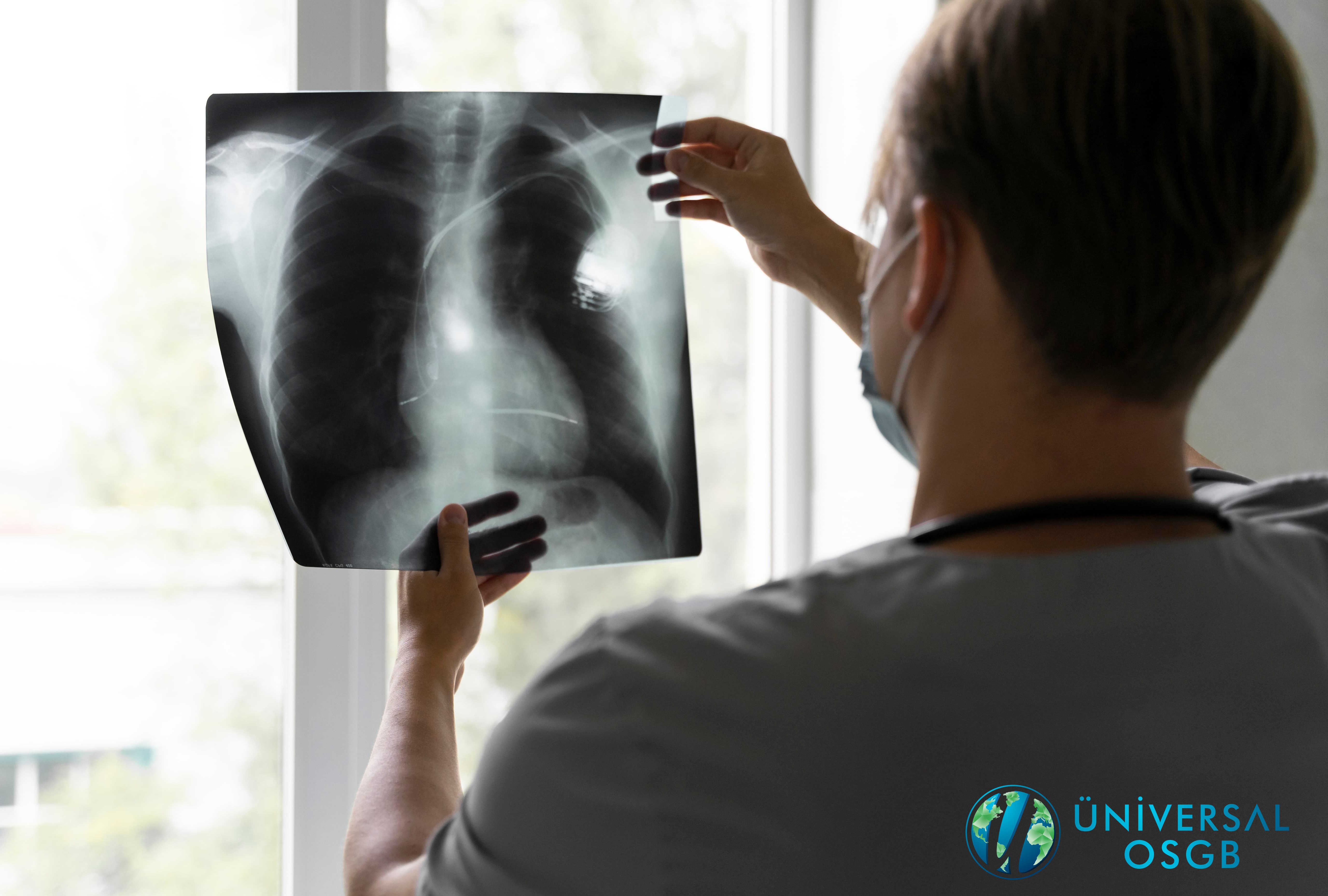
Lung radiography; It is a method used to determine relevant health problems by taking images of the heart, lungs, blood vessels, respiratory tract, and chest and spine bones. A chest x-ray can also show fluid in or around the lungs or air surrounding the lungs.
Lung radiography provides detailed imaging in a short time. Based on the images obtained, it can be determined whether there are health problems, a diagnosis can be made and the necessary treatment planning can be made.
Lung radiography is a diagnostic tool, especially in smokers and individuals with other respiratory disorders, and in employment examinations. It is frequently used as. However, repeated chest radiography may increase the individual's radiation dose, especially in the chest area. This may increase the risk of radiation-related diseases such as lung cancer. Additionally, unnecessary chest radiographs can increase the cost of healthcare and create unnecessary anxiety and stress.
Dose-Effect Relationship of Radiation and the Importance of Minimum Effective Dose
The effects of radiation on health are related to the dose. The dose-escalation relationship means that health risks increase with increasing radiation dose. Therefore, radiation doses used in medical imaging procedures should be as low as possible. The minimum effective dose is the lowest level of dose required for diagnosis and treatment purposes and is important to protect the health of the patient.
Today, it is estimated that 1-2% of all cancers in the world may be due to radiological examinations. In fact, in daily life, hair Radiation can be received from many sources, from drying machines to mobile phones and computers. However, the effects associated with these generally remain at relatively low levels.
The unit frequently used in radiation measurements is called Millisvert-mSv. For example, the radiation dose (0.02 mSv) exposed to a one-way chest x-ray is half of the radiation exposure (0.04 mSv) during a 6-hour plane journey. On the other hand, in lung tomography, this dose is at the level of 5-7 mSv. Although the threshold radiation level at which the risk of cancer increases is not clearly known in studies, it is generally reported that the relationship between cancer risk and radiation dose is directly proportional. Another risk that comes with lung tomography is radiation exposure to thyroid and breast tissue. Although various precautions can be taken to protect the individual from radiation during CT examinations, unnecessary tomography scans may cause the person to receive extra radiation. For this reason, it is necessary to avoid having more than 2 tomography scans a year, except in cases of absolute medical necessity, and to obtain information from a Chest Diseases Specialist regarding medical necessity situations.
The harms of radiation from excessive chest radiographs on human health should be carefully evaluated. Unnecessary repeated imaging procedures should be avoided and radiation doses should be kept as low as possible. Additionally, careful evaluation must be made to ensure that the benefits of medical imaging procedures exceed their risks. This is important to protect the health of both the individual and the society.
This article addresses the issue from an academic perspective, focusing on the harms of radiation from excessive chest X-rays on human health. In the light of relevant research, it has been emphasized that radiation doses should be kept under control and unnecessary repeated imaging procedures should be avoided.
It is thought that training patients about radiation in medical imaging devices can be provided.
Üniversal OSGB
Occupational Safety and Worker Health Center